After proteins are made by ribosomes during translation (here is a previous post covering how proteins are made), they require many additional chemical modifications to fold into the right shape to perform their different functions. These changes are essential because even the smallest changes to structure can dramatically alter how a protein behaves in the cell.
These modifications, called post-translational modifications ( PTMs ), usually involve attaching one or more molecules to the R group of an amino acid via a covalent bond. In this post, we’ll explore how these modifications occur, some examples of the most common ones, and how errors in the process can lead to the rise of cancerous cells.
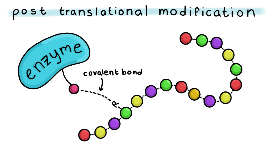
It is essential that these modifications occur because protein structure determines the function. PTMs help proteins fold into the right shape to carry out the different tasks needed in the cell.
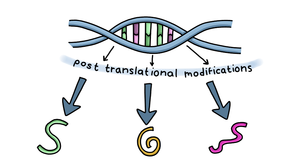
Scientists have identified around 200 PTMs in the cell. These different types of modifications help increase the diversity of the proteome, which is the complete set of proteins expressed by an organism. As a result, this diversity allows one gene to give rise to many different types of proteins depending on how the protein is modified after translation.
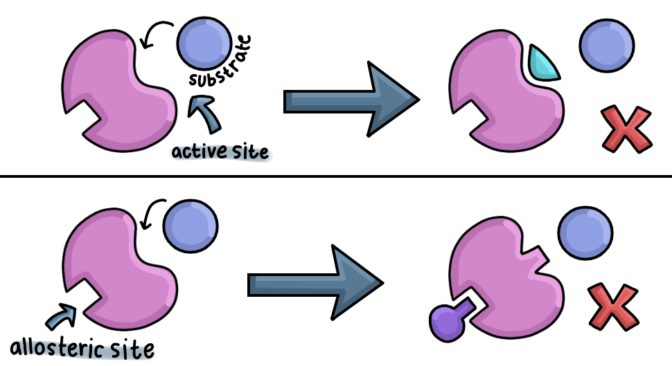
Post-translational modifications can occur:
- At or near the functional/ active site ( oetriosteric )
- Away from the functional site ( allosteric )
The functional/active site of a protein is responsible for binding substrates or carrying out its primary function. Modifications at the active site can directly affect protein activity, while allosteric modifications alter the protein’s shape indirectly and regulate activity more gradually.
Some PTMs are catalyzed by enzymes and are highly regulated, while others occur spontaneously. Spontaneous modifications can be either reversible or irreversible, although many spontaneous modifications are irreversible and can accumulate over time. Reversible modifications can be either removed via an enzyme or through the general instability of the bond.
Some common post-translational modifications include:
- Phosphorylation
- Methylation
- Acetylation
- SUMOylation
- Glycosylation
- Ubiquitination
All of these types of PTMS have different jobs to make sure that proteins fold to match their function. PTMs can occur in any step throughout the lifespan of a protein, allowing them to be regulated and repressed for different processes in the cell, like cell growth or division.
In this post, we’ll focus on 3 specific types of PTMS related to cancer, namely:
- Phosphorylaton
- Methylation
- Ubiquitination
We’ll also go into more detail on how cancer exploits the ubiquitin-proteasome system to promote uncontrolled growth and division.
Phosphorylation: Modifying Cell Signals
Phosphorylation is one of the most common and important PTMs. In this process, enzymes called protein kinases transfer a phosphate group from ATP onto a target protein.
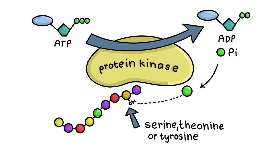
Protein kinases play key roles in cell signaling pathways. They can activate a protein, deactivate it, or allow it to pass signals to downstream proteins via phosphorylation.
Most phosphorylation occurs on the 3 amino acids:
- Serine
- Threonine
- Tyrosine
The enzyme attaches the phosphate group to the OH group on these amino acids. Different protein kinases target different amino acids. For example, serine/threonine kinases phosphorylate serine or threonine amino acids only.
Enzymes called phosphatases can reverse this reaction and remove the phosphate group from the target protein.
Ubiquitination: The Cell’s Protein Recycling System
This is another major PTM and is the most common PTM after phosphorylation. This modification attaches a small protein called ubiquitin to a target protein through a reversible covalent bond.
Ubiquitination is part of a larger process in the cell called the ubiquinin- proteasome system (UPS), which tags proteins to be degraded in the proteasome, and regulates the protein turnover in the cell.
There are 3 enzymes that facilitate this process:
- Ubiquitin activating enzyme (E1)
- Ubiquitin conjugating enzyme (E2)
- Ubiquinin ligase (E3)
First, E1 uses ATP and turns it into AMP to activate the carboxyl end of ubiquitin and attaches it to the cysteine residue in the active site of E1 through a thioester bond. This activated ubiquitin is then transferred to the cysteine amino acid of E2 via a transesterification reaction.
A transesterification reaction is where the R group of an ester is transferred to the R group of an alcohol, via the replacement of the OR group. E3 then transfers the ubiquitin from E2 to the lysine amino acid in the target protein via an isopeptide bond. This is a type of bond that forms between a lysine amino acid and a carboxyl group.
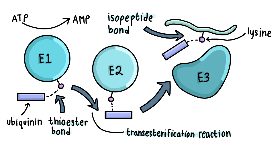
There are 8 different types of linkages for ubiquitination, each serving as a signal for different fates in the cell. For example, K48-linked polyubiquitin chains target proteins for degradation by the proteasome. Generally, 4 or more ubiquitin molecules must be present on the target protein for degradation.
The 26S proteasome is in charge of degrading the ubiquitinated proteins. The 20S core particle is made of alpha and beta rings, and there are 2 19S regulatory particles on the top and bottom of the proteasome used for recognition of the ubiquitinated protein.
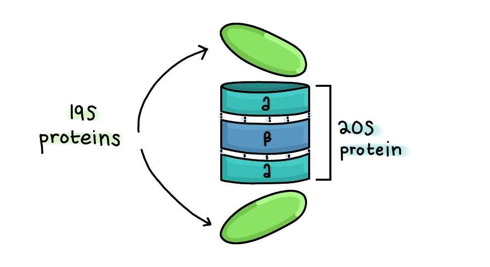
The target protein unfolds to its primary structure as it enters via hydrolysis of an ATP bound to the lid, and enters the protease site located in the B subunit. It is cleaved inside and leaves the proteasome as smaller proteins to be broken down into individual amino acids by other enzymes.
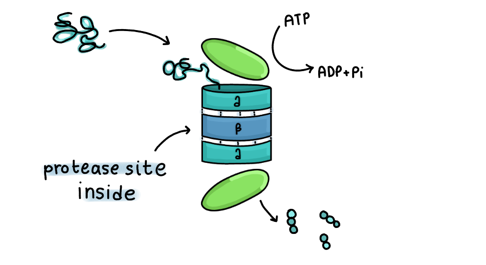
There is a special cleavage site inside the proteasome for isopeptide bonds to allow the cell to recycle the ubiquitin proteins to be used later, removing these molecules before degradation occurs.
Glycosylation: Adding Sugars for Structure and Function
In this PTM, one or more sugar molecules ( usually a monosaccharide or oligosaccharide) are covalently attached to a protein or lipid via glycosidic bonds.
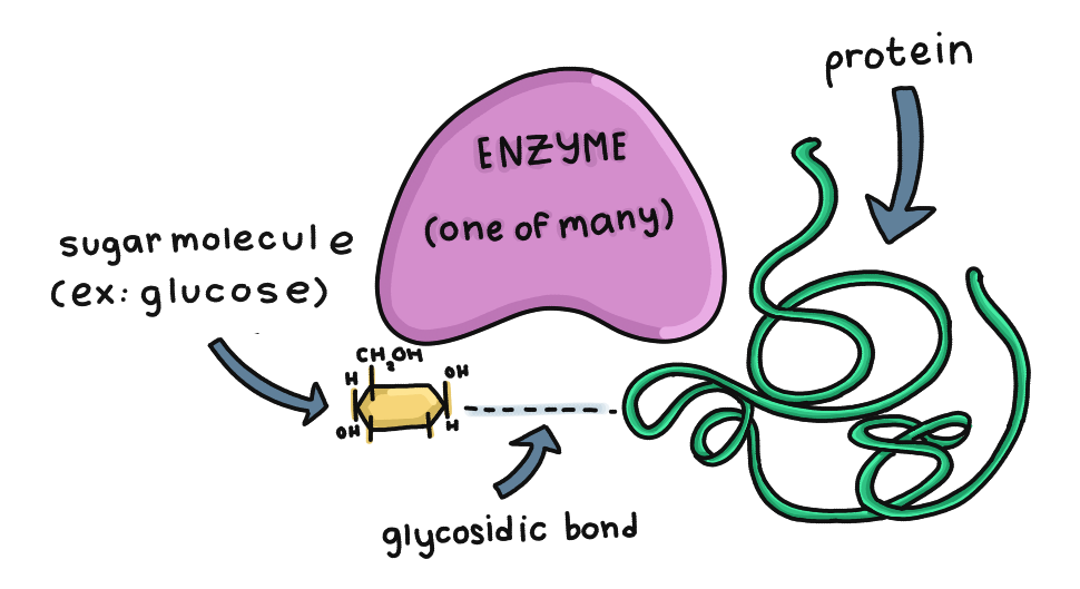
The 2 most common types of glycosylation are:
- N linked
- O linked
In N-linked glycosylation, sugar molecules are attached to the Asparagine (Asn) amino acid in the protein. An enzyme called oligosaccharyltransferase transfers a preassembled oligosaccharide from a lipid carrier into the protein. Lipid carriers are molecules that aid in making the sugar and then transferring it for glycosylation.
This process begins in the rough endoplasmic reticulum (RER) and continues through the Golgi apparatus.
In O-linked glycosylation, enzymes called glycotransferases attach sugar molecules to the hydroxyl group in a serine or threonine amino acid in the protein. Unlike N-linked glycosylation, this process primarily occurs in the Golgi apparatus.
How Cancer Exploits the Ubiquitin Pathway
Cancer cells can exploit the process of ubiquination and the ubiquinin- proteasome system to allow uncontrolled growth and survival. Many different processes around the cell are powered by this system, and the dysregulation can cause tumor formation.
Here are some examples of how the ubiquitin pathway is dysregulated in cancer:
- E1 Enzymes
- These enzymes help E3 ligases for DNA damage repair. They also control cell cycle regulation and apoptosis via ubiquitin. The dysregulation of the enzyme can lead to a defective DNA repair mechanism and increase mutation rates, contributing to cancer development.
- E2 Enzymes
- E2 enzymes also partner with E3 ligases to regulate proteins involved in the cell cycle.
- One example is UBE2C, which regulates mitosis by the ubiquitination and degradation of cell cycle regulators in mammal cells. It is overexpressed in cancer and can cause unchecked cell cycle progression and promote the growth of tumors.
- E3 Enzymes
- There are many different types of E3 ligases in the mammalian genome. One specific example is the HECT ligase, where some members of the subfamily regulate oncogenic and tumor suppressor genes. The dysregulation of the ligase can lead to the progression of cancer by targeting proteins that normally stop cancer growth.
Here are some other examples of how the dysregulation of ubiquitination in the cell in general can accelerate cancer growth:
- P53 Proteins
This is an example that we have talked about in a previous blog post. These are a type of tumor suppressor protein whose levels are tightly controlled by the MDM2 E3 ligase enzyme, which binds to the protein and marks it for degradation in the proteasome via ubiquitination keeping levels low.
If the protein accumulates in healthy cells, it can lead to apoptosis. In response to DNA damage, kinases like ATM phosphorylate both p53 and MDM2, disrupting their interaction and preventing ubiquitination. This causes the cell to activate DNA repair pathways or trigger apoptosis if the damage is too severe. When MDM2 is overexpressed in some cancers, it can lead to excessive degradation of p53, allowing the cell to escape apoptosis.
- Cyclins and Cyclin-Dependent Kinases
Cyclins and cyclin-dependent kinases (CDKs) regulate progression through the cell cycle.
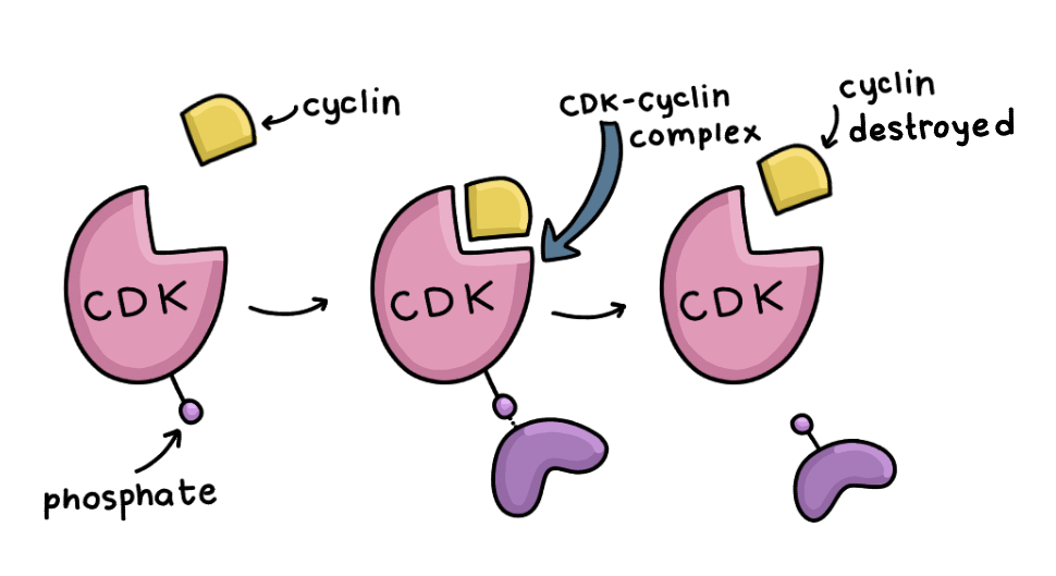
Cyclins are proteins that help regulate the cell cycle. Ubiquitination helps regulate the timely degradation of cyclins and allows the cell to transition between cell cycles. One example of this is the degradation of Cyclin B, which allows the cell to exit mitosis and go to the G1 phase.
The overexpression of certain kinds of cyclins can cause uncontrollable cell division. 2 examples include cyclins D and E, which allow the cell to skip essential checkpoints before dividing.
Ubiquitination also allows the degradation of inhibitors of CDK complexes at the right time and prevents unchecked cell cycle progression in normal cells. In cancerous cells, however, the process destroys them at the wrong time and allows the cells to grow and divide uncontrollably.
New Cancer Therapies Targeting Protein Regulation
Researchers are currently working on developing therapies that target the UPS in cancer cells.
Here are 2 of the most common therapies:
- Proteasome inhibitors
- Deubiquitination inhibitors
Proteasome inhibitors work by blocking the proteasome’s ability to break down proteins. They are designed to make the cell accumulate misfolded and damaged proteins, triggering apoptosis. However, these drugs can also affect healthy cells, leading to side effects and toxicity for the patient.
Another emerging therapy is deubiquitination inhibitors, which prevent the removal of ubiquitination proteins from their target proteins. Oncoproteins can be abnormally activated, and the removal of ubiquitin proteins leads to their stabilization and the inactivation of tumor suppressors. These drugs help prevent the stabilization of oncoproteins in the cell and allow for their degradation.
Conclusion
Post-translational modifications are important for allowing the cell to diversify the different proteins it produces. There is a wide range of different PTMs, all aimed at helping proteins specialize in whatever their specific jobs are. Cancerous cells take advantage of these processes, such as the ubiquitin pathway, which causes widespread dysregulation in the cell and allows the cell to grow and divide faster while evading the body’s defenses.
If you learned anything, make sure to send us a message on our Instagram (@whencellsresist). We love hearing back from our readers!
All original insights and illustrations are my own. This content is not intended as medical advice.
Drawings are simplified for illustrative purposes and may not be exact representations of the subjects
Sources
https://pmc.ncbi.nlm.nih.gov/articles/PMC10546021
https://www.news-medical.net/health/-What-is-Protein-Phosphorylation.aspx
https://www.creative-proteomics.com/blog/protein-phosphorylation.htm
https://www.youtube.com/watch?v=_AwXhA_gncc
https://www.youtube.com/watch?v=0VFF7-GyX00
https://www.youtube.com/watch?v=phCeSvypNLI
https://www.youtube.com/watch?v=qFqbueKBd8w
https://www.youtube.com/watch?v=xHpQuV80xhs
https://www.creative-proteomics.com/resource/ubiquitination-protein-degradation-in-cancer.htm
https://link.springer.com/article/10.1186/s12943-024-02046-3
https://pubmed.ncbi.nlm.nih.gov/40537889
https://www.youtube.com/watch?v=D4QXR8Exzjo
https://www.youtube.com/watch?v=kI_JfNduhYg